Quick Answer
- DVT Treatment Goals: Preventing clot growth and reducing the risk of new clot formation.
- Diagnosis: Wells score followed by D-dimer testing and confirmation with compression duplex ultrasound.
- Clot Stabilisation: Anticoagulant therapy is the primary treatment for deep vein thrombosis.
- Clot-Dissolving Treatment: Thrombolytics or clot-busters are used in severe cases to dissolve blood clots.
- Circulation Support: Compression stockings help reduce swelling and improve venous blood flow.
- Prevent Pulmonary Embolism: Inferior vena cava (IVC) filters are used in patients who cannot receive anticoagulant therapy to stop clots from reaching the lungs.
- Treatment Duration: Most DVT treatment plans last at least 3 months, depending on the cause of the clot and individual risk factors.
DVT treatments focus on preventing the clot from growing, reducing the risk of pulmonary embolism (PE) and restoring venous circulation.
The management of DVT includes anticoagulant therapy as the first line of treatment, with compression therapy and lifestyle adjustments that support recovery. If there are large clots, minimally invasive procedures may be used to remove or dissolve them & save the limb.
Early diagnosis and timely leg blood clot treatment significantly reduce complications like PE and improve long-term vascular health.
DVT: Clinical Definition
According to NCBI, Deep vein thrombosis is a condition where a blood clot forms in the deep veins, most commonly of the legs or pelvis, but can also occur in the arms, mesenteric and cerebral veins.
Symptoms of DVT
Symptoms of DVT include sudden pain with swelling in the thigh or calf region, only in one leg. In most cases, the condition develops gradually and may show minimal or no symptoms.
Common DVT symptoms include:
- Swelling in one leg, usually affecting the calf or thigh
- Pain or tenderness in the leg, particularly during walking or standing
- Warmth and redness over the affected vein
- Visible or enlarged veins caused by impaired venous circulation
- Mild or no symptoms in some individuals, which can delay diagnosis.
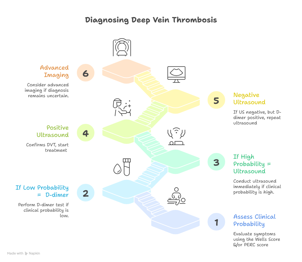
How is Deep Vein Thrombosis Diagnosed?
As per NICE guidelines, diagnosis of deep vein thrombosis begins with clinical probability assessment using the Wells score and, where required, the Pulmonary Embolism Rule-out Criteria (PERC).
This is followed by D-dimer testing and confirmation with compression duplex ultrasound, ideally performed within 4 hours. If initial results are negative but clinical suspicion remains high, repeat ultrasound after 6–8 days is recommended.
Although contrast venography was historically considered the gold standard, duplex ultrasound is now the preferred first-line test because it is non-invasive, accurate, and safe.
Accurate diagnosis is essential because untreated DVT can progress to PE or chronic DVT/post-thrombotic syndrome (PTS).
Clinical Assessment and Risk Evaluation
- Detailed medical history and physical examination focus on symptoms like leg swelling, pain or redness, along with risk factors such as recent immobility or surgery.
- Risk scoring tools, such as the Wells score, estimate the likelihood of DVT and guide further testing.
D‑Dimer Blood Test
- A D‑dimer test measures the clot breakdown fragments in the blood.
- Low levels of D‑dimer in a low-probability setting can effectively rule out DVT, reducing the need for imaging.
- However, D‑dimer is not specific to clot presence and may be elevated in many conditions (e.g., infection, inflammation, pregnancy), so it cannot confirm DVT alone.
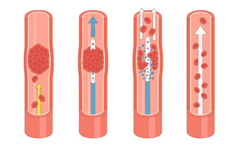
Compression (Duplex) Ultrasound
- Non-invasive imaging that uses sound waves to visualise blood flow and vein compressibility.
- In a normal vein, gentle pressure from the ultrasound probe causes the vein to flatten, while if a vein fails to compress, it suggests a clot.
- This test successfully identifies most clots in large leg veins.
Additional Imaging
- If ultrasound results are inconclusive, further imaging is used.
- Options include venography (an X-ray with contrast) and magnetic resonance imaging (MRI), which can show clots not visible on ultrasound.
- Further imaging may be required to detect underlying abdominal pathology that could lead to atypical DVT.
Integrating Tests for Accurate Diagnosis
- Combining clinical evaluation with laboratory and imaging tests improves diagnostic accuracy.
- Early diagnosis allows timely initiation of DVT treatment to prevent clot extension and reduce risks of PE or PTS.
Common Treatments for Deep Vein Thrombosis
The primary goal of DVT treatment is to prevent clot extension, reduce the risk of pulmonary embolism and protect long-term venous function.
Anticoagulant Therapy
- Anticoagulants form the foundation of DVT treatment, preventing clot extension and new clot formation.
- They allow the body’s natural processes to gradually break down the clot.
- Early initiation significantly reduces the risk of PE or recurrence.
- Treatment continued for at least 3 months, with longer duration in high-risk patients.
Compression Therapy
- Compression stockings apply graduated pressure, highest at the ankle, to support upward blood flow.
- They help reduce swelling, discomfort, and heaviness in the affected leg.
- Regular use may lower the risk of chronic DVT/PTS.
- Stockings are recommended for several months during recovery.
Catheter-Directed Thrombolysis
- A minimally invasive procedure used in selected patients with large or severe clots.
- An ultrasound-guided catheter is inserted into the affected vein to deliver clot-dissolving medication called ‘Clot Busters’ directly to the blocked area.
- Indicated for large clots in the upper thigh veins (especially in younger patients), clots in the veins of shoulder region, or in severe cases where the clot must be dissolved quickly to protect the li
Inferior Vena Cava (IVC) Filters
- An IVC filter is a small device placed in the Inferior Vena Cava, the largest vein in the abdomen that carries blood from the lower body to the heart.
- The filter works by trapping clots before they reach the lungs, helping prevent PE.
- It is generally used when anticoagulant therapy is not suitable due to bleeding risk or other medical concerns.
- Many filters are temporary and may be removed once the risk of clot migration decreases.
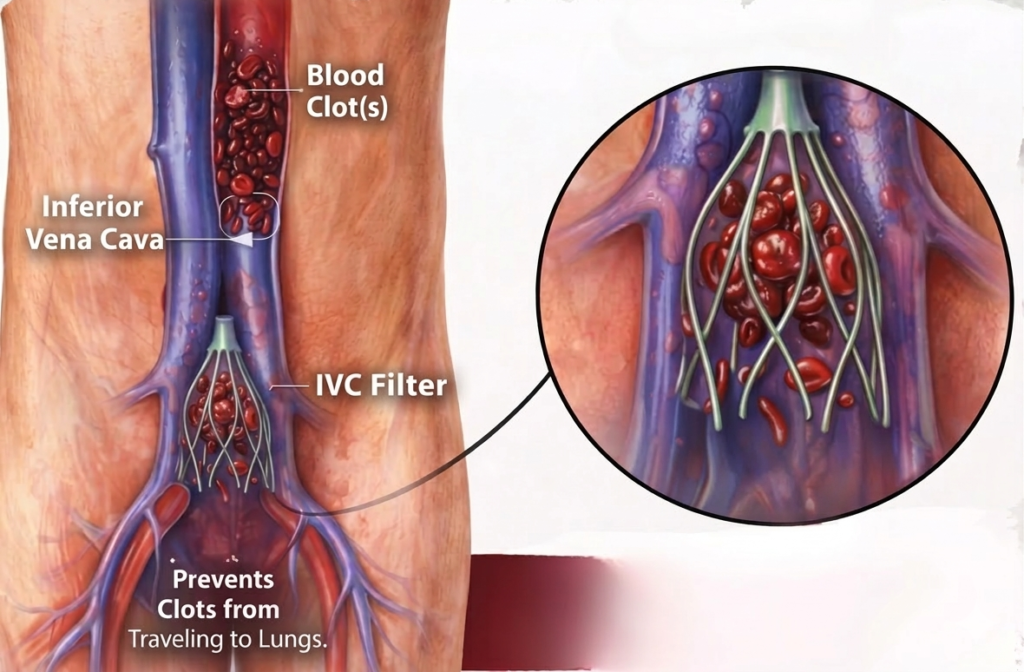
Note: The decision to remove IVC filters is based on clinical assessment and imaging results. IVC filter retrieval is a separate procedure performed using a catheter under light sedation.
Lifestyle and Recovery Measures
These measures support recovery, prevent new clot formation and improve long-term outcomes.
- Regular walking and leg movement to stimulate circulation.
- Avoid prolonged sitting or immobility to reduce clot risk.
- Maintain hydration, healthy weight and active lifestyle.
Complications Related to Deep Vein Thrombosis
- Pulmonary embolism (PE): A life-threatening condition caused by a clot travelling to the lungs.
- Post-thrombotic syndrome (PTS): Chronic pain, swelling and skin changes due to long-term vein damage.
- Recurrent DVT: Increased risk in patients with persistent underlying factors.
- Chronic venous insufficiency (CVI): Long-term impairment of venous circulation in the affected limb.
Take Home Message
DVT treatment aims to prevent clot growth, reduce the risk of pulmonary embolism & complications and restore healthy circulation.
Diagnosis relies on clinical risk assessment, D-dimer testing, and confirmation with compression duplex ultrasound, allowing timely identification of the clot. Treatment prioritises anticoagulant therapy first to stabilise the clot, compression therapy to improve circulation, and minimally invasive procedures in selected cases, along with lifestyle measures that support recovery.
Early recognition of symptoms such as unilateral leg swelling, pain, warmth, or redness is important because untreated DVT can lead to life-threatening complications like pulmonary embolism or long-term vein damage known as chronic DVT/Post‑Thrombotic Syndrome.
FAQs
How long is recovery from a DVT?
DVT recovery timeline
- 1–2 weeks: Pain and swelling begin to improve after starting blood thinners; light walking is encouraged.
- 2–6 weeks: Many patients return to most normal activities, though mild swelling or discomfort may persist.
- 6 weeks – 3 months: Symptoms resolve for most patients as the clot stabilises and circulation improves.
- Beyond 3 months: Some patients require continued monitoring or extended treatment depending on individual risk factors.
Factors affecting recovery from DVT:
- Size and location of the clot
- How early treatment is started
- Presence of underlying risk factors (cancer, surgery, or prolonged immobility)
- Patient age and overall health
- Adherence to anticoagulant therapy
- Follow-up care
Recovery may take longer in patients if complications such as PTS or PE dev
Do compression stockings help with deep vein thrombosis?
Yes, compression stockings are recommended as part of DVT treatments. They apply graduated pressure to the legs, improving venous blood flow and reducing swelling.
Compression therapy can also help prevent post-thrombotic syndrome, a long-term complication that may occur after a clot. Proper fitting and medical guidance are important to ensure effective compression therapy.
How to prevent deep vein thrombosis at home?
Preventing DVT focuses on improving circulation and reducing clot risk.
Helpful prevention strategies include:
- Regular movement and walking, especially during long travel
- Staying well hydrated and maintaining a healthy weight
- Avoiding smoking and prolonged inactivity
- Following medical advice for preventive therapy if at high risk
These deep vein clot prevention strategies help protect vascular health and reduce the likelihood of future clot formation.