
GI Bleed
GI bleed, or gastrointestinal bleed, is basically bleeding from the digestive system, and it is never something to wait out. It can be sudden and heavy, or slow and silent, and in both cases, it leads to blood-stained stools or vomit along with a sudden drop in haemoglobin.
Every GI bleed ultimately comes from a blood vessel and is mostly managed with medication or endoscopy. When that doesn’t work or the bleed it out of reach, a vascular specialist should step in.
Read on to learn what causes GI bleeding and how it is treated.
What Is a GI Bleed?
A gastrointestinal (GI) bleed is bleeding from any part of the digestive tract, from the food pipe and stomach to the intestines, colon, rectum or anus.
Based on where the bleeding starts, it can be of 2 types:
- Upper GI bleed, which comes from the food pipe, stomach or first part of the small intestine (duodenum).
- Lower GI bleed comes from the rest of the small intestine, colon, rectum or anus.
Upper bleeds are more common and usually more serious because blood loss can be rapid.
GI Bleed starts when a blood vessel in the gut wall erodes, ruptures or is abnormally fragile or swollen. The cause may be an ulcer, a tumour or inflamed tissue.
GI Bleed Symptoms
Vomiting Blood
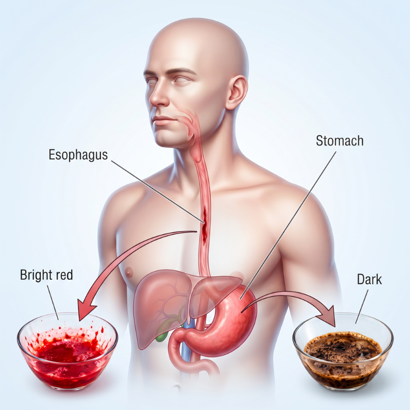
Bright red blood, or dark material that looks like coffee grounds in your vomit, indicates an upper GI bleed.
Blood in Stools
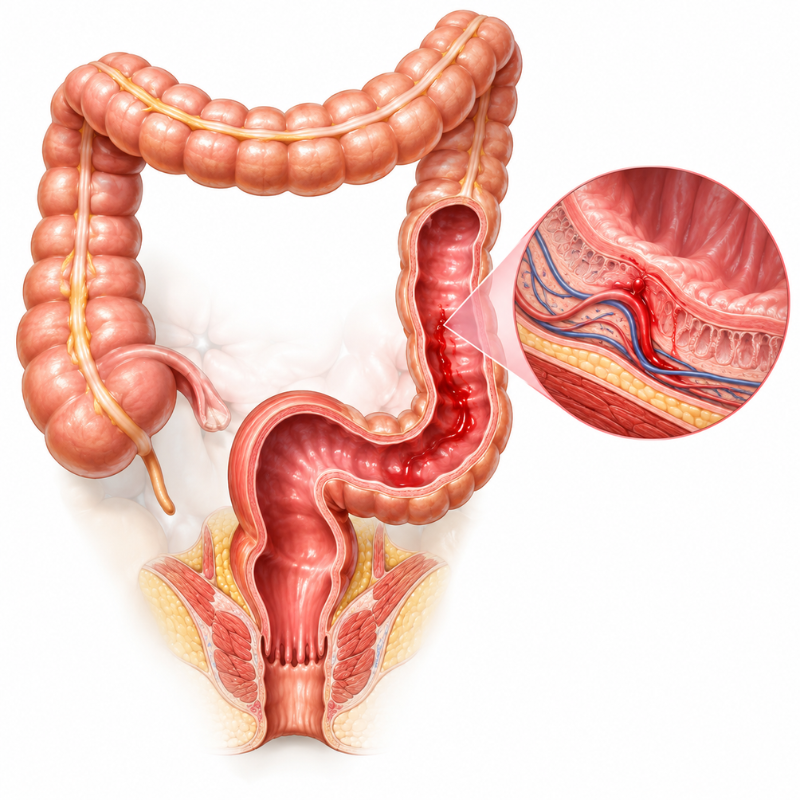
Bright red or maroon blood in the stools comes from the rectum or anus, or slightly higher areas in the lower gut.
Sticky, dark-coloured stools (melaena) usually suggest bleeding higher up in the tract.

Dizziness
Tiredness & Pallor
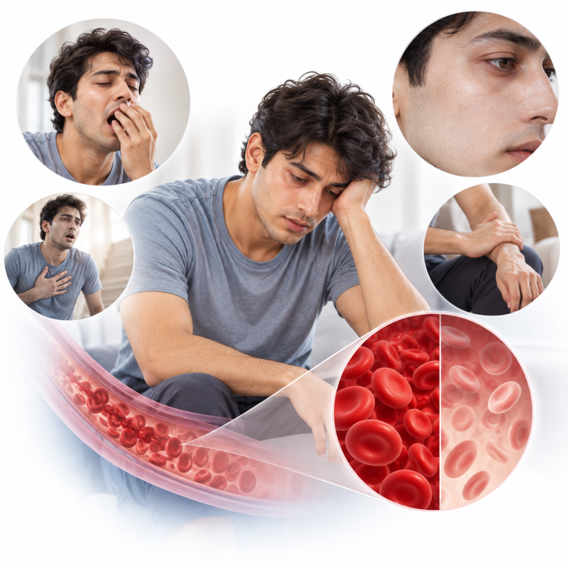
Slow, hidden bleeding shows as unexplained tiredness, pale skin, weakness, breathlessness and low haemoglobin.
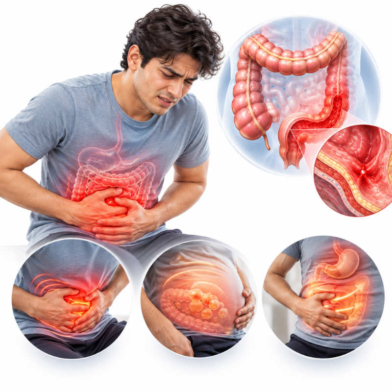
Abdominal Pain
Burning or cramping pain, bloating, or fullness can accompany the bleed.
If you vomit blood, pass black or bloody stools, or feel faint, go to the nearest emergency department immediately.
GI Bleed Causes
Upper GI Bleed
Peptic Ulcers

Peptic Ulcers
Open sores in the stomach or duodenum, often linked to H. pylori infection or regular painkiller use, are the most common cause of upper GI bleeding.
Painkillers (NSAIDs)
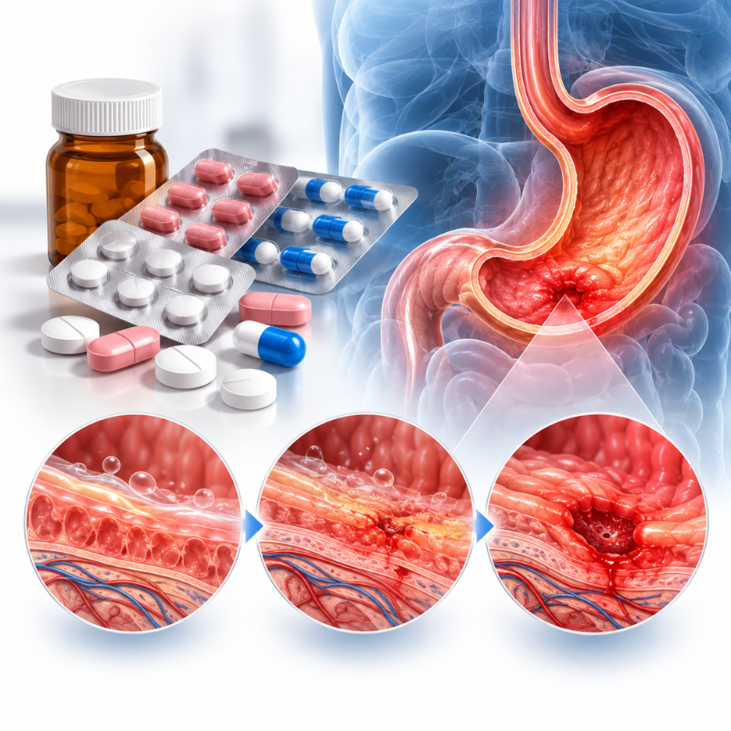
Painkillers (NSAIDs)
Diclofenac, ibuprofen and aspirin weaken the stomach’s protective lining, especially with long-term or unsupervised use.
Oesophageal and Gastric Varices
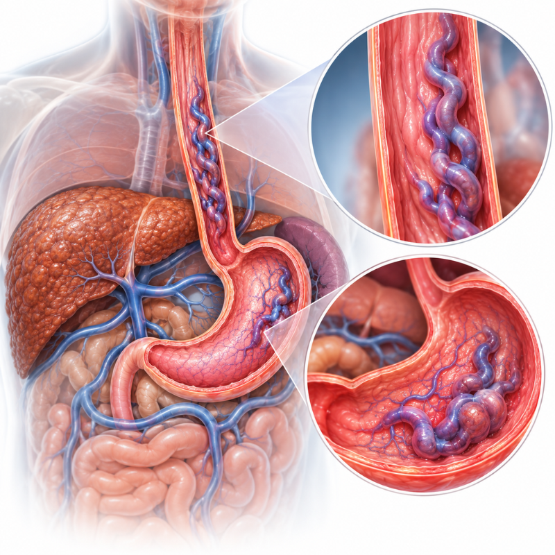
Oesophageal and Gastric Varices
Swollen veins in the food pipe or stomach are caused by raised pressure, as in liver disease. These can bleed heavily and suddenly.
Gastritis and Erosions
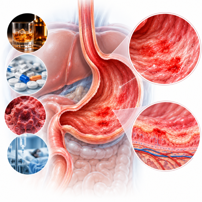
Gastritis and Erosions
Lower GI Bleed
Piles and Anal Fissures
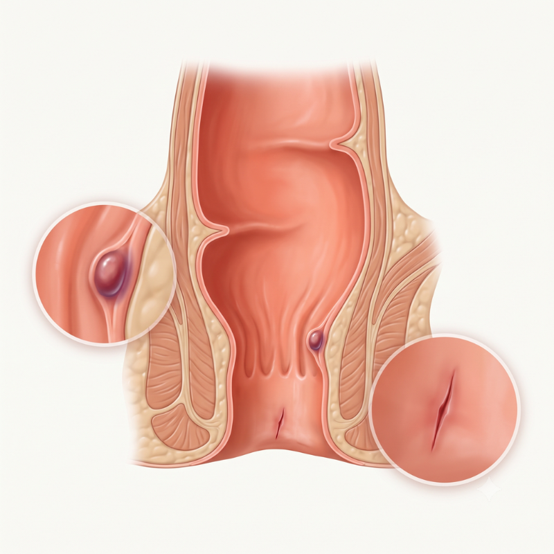
Piles and Anal Fissures
Diverticular Disease
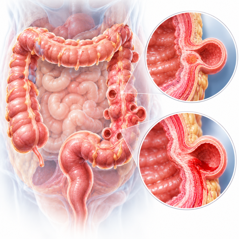
Diverticular Disease
Angiodysplasia
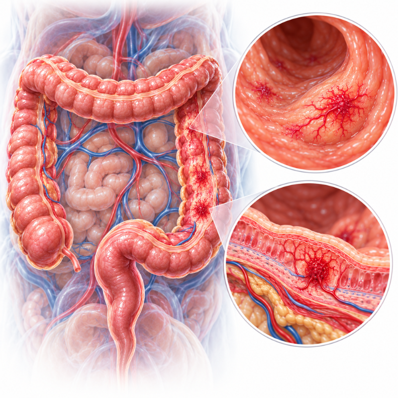
Angiodysplasia
Polyps and Tumours
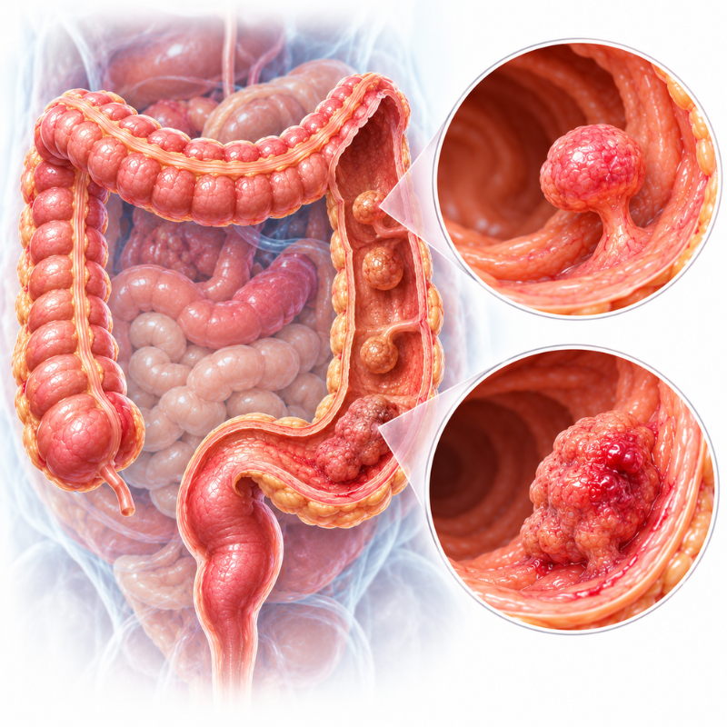
Polyps and Tumours
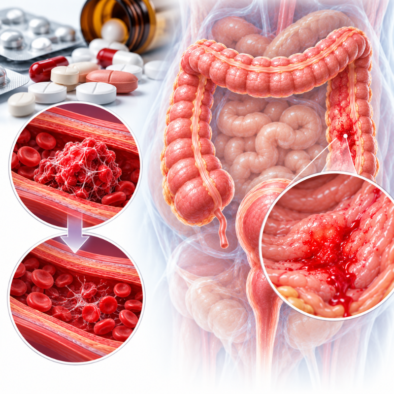
Blood Thinners
Blood Thinners
Anticoagulant and antiplatelet medicines do not cause the bleed themselves, but can make an existing one heavier and harder to stop.
GI Bleed Diagnosis
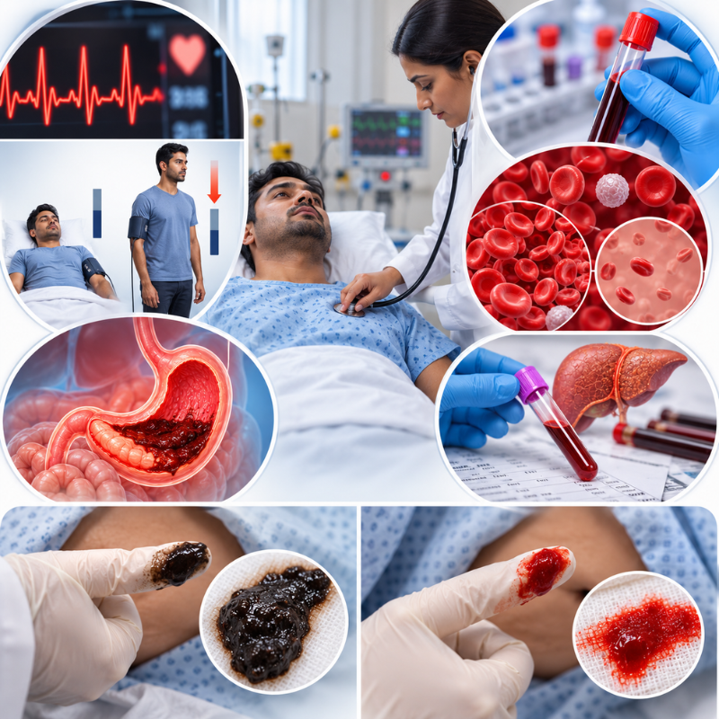
Clinical Exam and Blood Tests
- Fast pulse and low blood pressure, often with a drop in BP on standing
- Low haemoglobin
- Raised blood urea, often seen in upper GI bleeds
- Prolonged clotting time (raised INR) in liver disease or with blood thinners
- Black stool or fresh blood on rectal examination
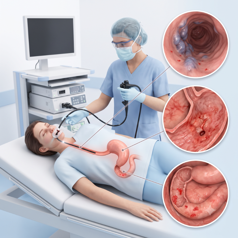
Upper GI Endoscopy
A thin camera is passed through the mouth to examine the food pipe, stomach and duodenum.
It is the first test for upper bleeds and can often treat the source at the same time.
- Ulcer with an exposed vessel, active oozing or a clot
- Varices (swollen veins) with red marks or active bleeding
- Gastritis, erosions, or a growth in the food pipe or stomach
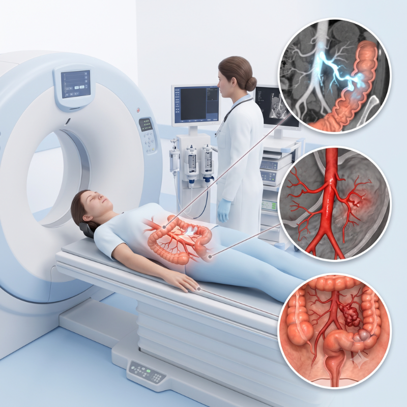
CT Angiography
A rapid scan that uses contrast dye to show where blood is leaking from a vessel.
It is especially useful when endoscopy cannot find the source or the bleeding is too heavy to see clearly.
- Contrast leaking into the bowel, which confirms active bleeding and shows its site
- The artery feeding the bleed
- Underlying cause, such as a growth, diverticula or an abnormal vessel
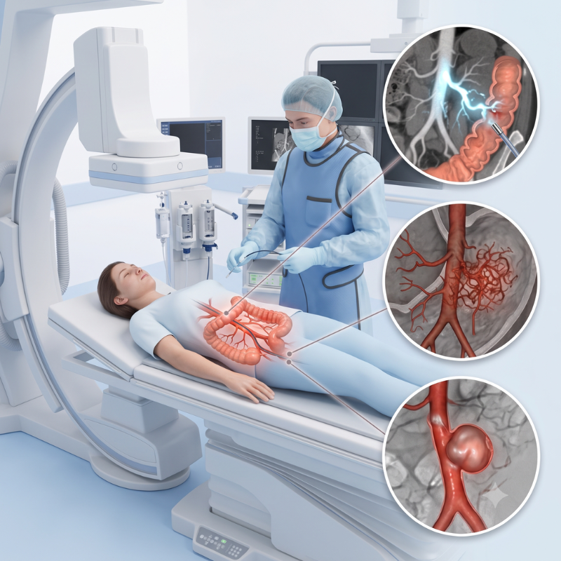
Catheter Angiography
GI Bleed: Treatment
Treatment depends on the cause, site, and severity of the bleeding.
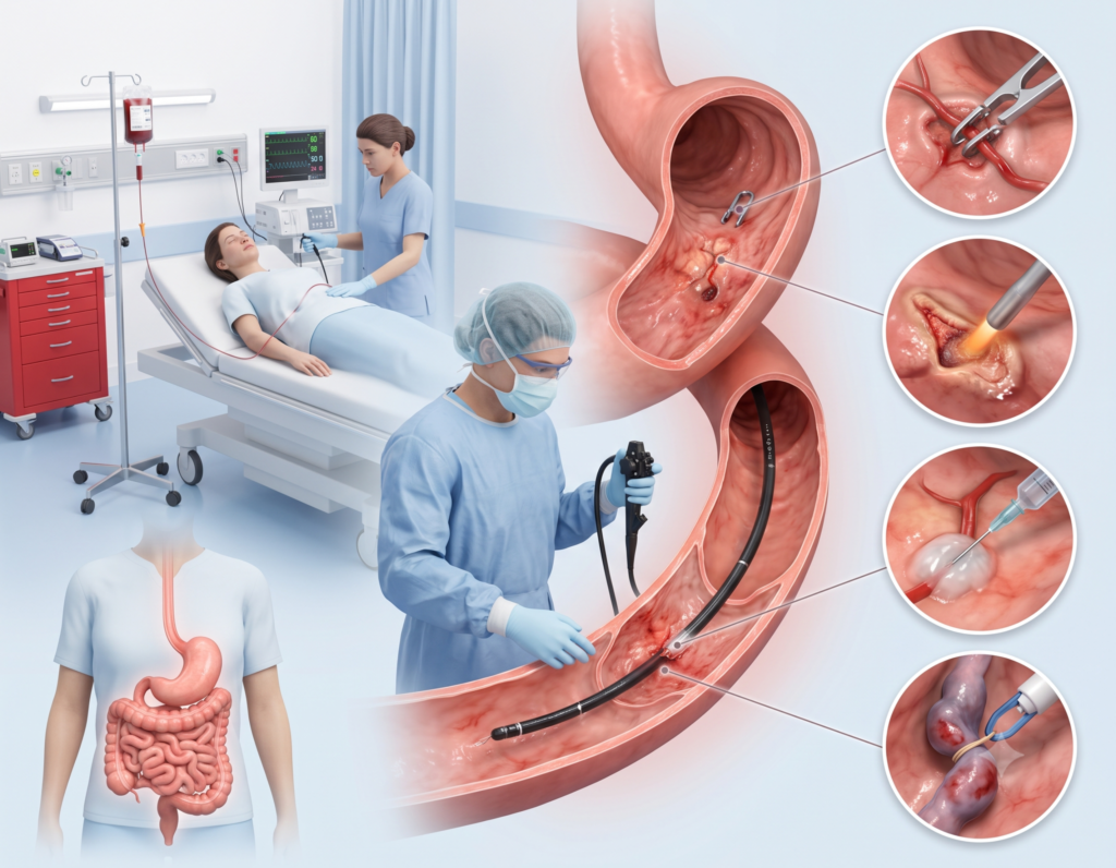
Emergency stabilisation is the first step, and endoscopy with clips, heat therapy, injection therapy or ligation applied directly at the bleeding site helps control most bleeds.
When these efforts fail, vascular surgeons employ the following procedures to control the bleeding
Angiographic Embolisation

Angiographic Embolisation
When bleeding continues after endoscopy, keeps returning, or the source cannot be reached, the bleeding vessel is blocked from the inside through a thin catheter, using coils, particles or medical glue.
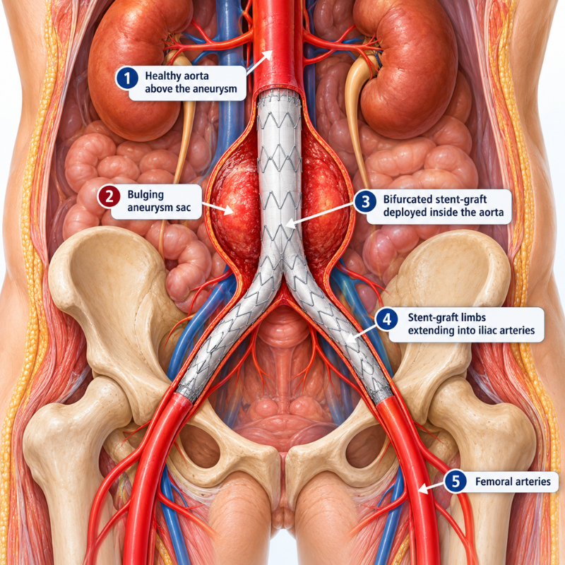
Stent Grafts and Repair of Bleeding Vessels
Stent Grafts and Repair of Bleeding Vessels
Some GI bleeds come from a damaged artery rather than the gut lining, and need the vessel itself repaired.
- Pseudoaneurysm: A weakened, bulging wall of an artery near the stomach, pancreas or liver (often after pancreatitis or surgery) that ruptures into the gut may be treated with coiling or a covered stent.
- Aortoenteric fistula: an abnormal link between the aorta and the bowel, either from an aortic aneurysm or after previous aortic surgery. An endovascular stent graft (EVAR) can stop the bleeding as a life-saving measure.
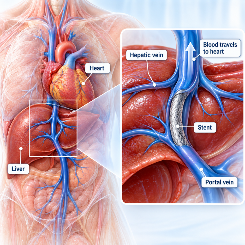
TIPS (Transjugular Intrahepatic Portosystemic Shunt)
TIPS (Transjugular Intrahepatic Portosystemic Shunt)
For bleeding venous clusters (varices) in liver disease that are uncontrolled or keep returning, TIPS may be recommended.
TIPS (transjugular intrahepatic portosystemic shunt) is a channel placed inside the liver to lower the pressure in the bleeding veins
GI Bleed Results: Before & After
Discover how minimally invasive treatments help relieve pain and improve the appearance of legs.
Comming Soon
Resources
Watch videos to understand how varicose vein treatments work and what to expect during recovery.
Read More
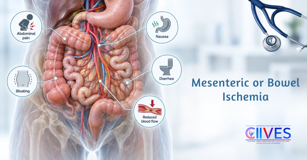
Mesenteric or Bowel Ischemia
Mesenteric ischemia (MeI): Occurs when reduced blood flow limits oxygen delivery to the intestine and surrounding organs, causing tissue injury and bowel necrosis.

A Curious Case of Fever & an 6-hour Race Against Time: Infected Aortic Aneurysm
An aortic aneurysm occurs when a weakened section of the aortic wall balloons outward. When infection reaches the vessel wall, however, it can cause inflammation and rapidly weaken the aorta.

Mesenteric Artery Bleeding or GI Bleed in Vascular Surgery
Mesenteric artery bleeding occurs when an artery supplying the intestines or one of its branches ruptures, leaks or tears.